Understanding the Bladder: Anatomy of the Urinary Tract
Reviewed by: HU Medical Review Board | Last review date: September 2017. | Last updated: April 2024
The urinary bladder, usually just called the bladder, is a major part of the body's urinary system.1-3 It is a hollow organ made mostly from muscle. The bladder's job in the urinary system is to store the urine produced by your body until it is released when you urinate.
How does the bladder store urine?
When it is empty, an adult's bladder is about the size and shape of a pear. The bladder is very flexible, like a balloon, which can expand and get bigger when it is full of urine. In most adults, the bladder can expand to hold about one pint of urine before they feel the urge to urinate. When the bladder is emptied after urinating, it shrinks back to its empty size.
Understanding the the urinary system
The body's urinary system is also called the 'renal' system, which comprises several different organs.1-3 The urinary system's job is to filter out waste and extra water from the blood and remove it from the body in the form of urine. The major organs that make up the urinary system are:
- Two kidneys
- Two ureters
- One bladder
- One urethra
The upper and lower urinary tracts
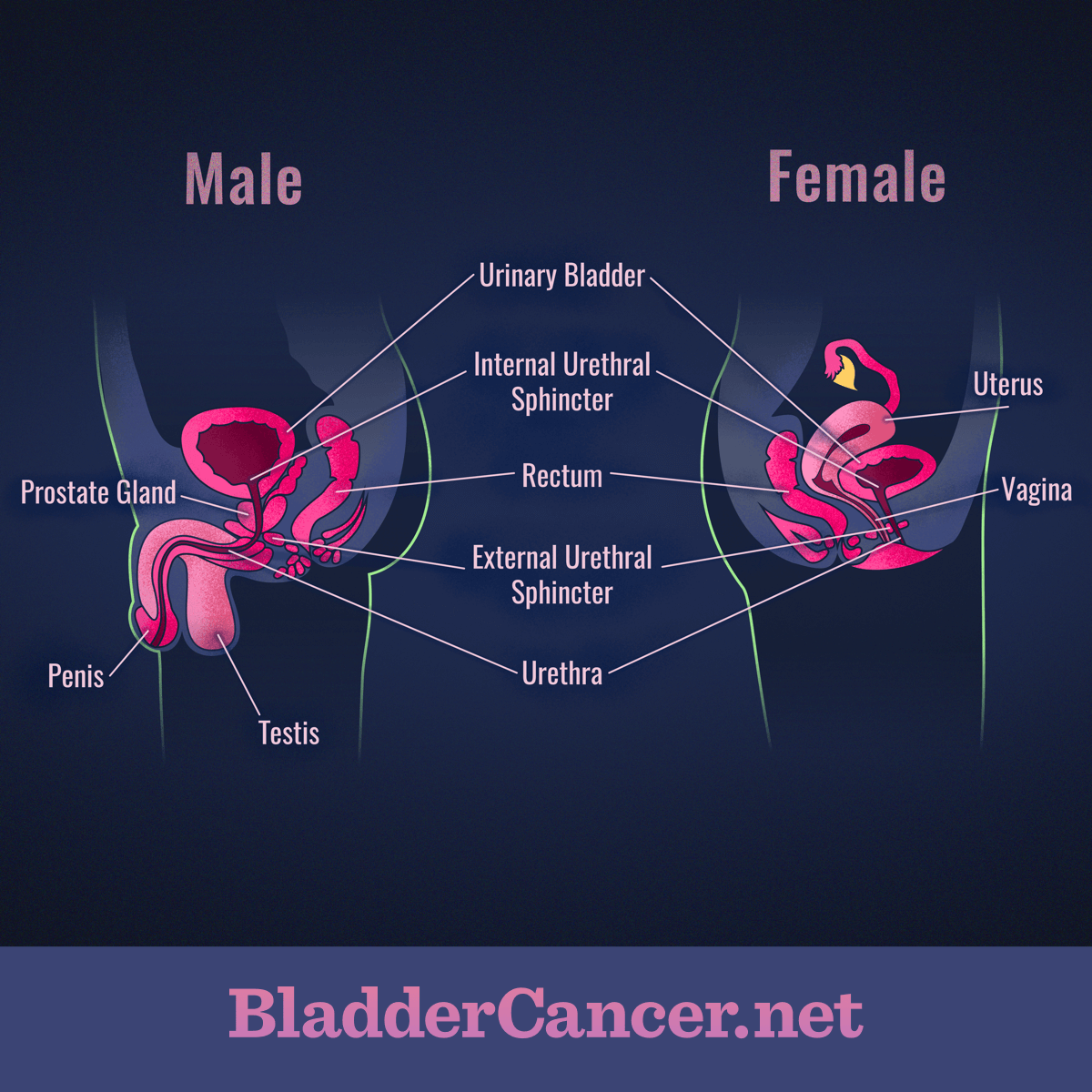
The urinary system has two parts: the upper and lower urinary tract. Together, the two kidneys and two ureters make up the upper urinary tract. The lower urinary tract contains the bladder and the urethra. Men and women have the same upper urinary tract, but their lower urinary tracts are different.
Figure 1. Comparison of male and female urinary systems
How does each part work?
In the upper urinary tract, the two kidneys work separately to filter your blood by removing waste products and extra water that have entered your bloodstream, mainly through digesting foods and drinks. Urine is made up of this filtered waste and water. The kidneys constantly work to filter your blood. As the kidneys produce urine, it is stored in a small area called the renal pelvis for a short time in the middle of each kidney. The renal pelvis inside each kidney is connected to the bladder by a long, thin tube called a ureter. In adults, each ureter is about 12 inches long. The renal pelvis acts like a kind of funnel. It regularly tightens up and sends the urine collected in the renal pelvis through the ureter and into the bladder.
The bladder is located in the pelvis, just behind the pubic bone in the lower urinary tract. When the bladder is full of urine from the kidneys, you feel the urge to urinate. During urination, the muscles in the bladder tighten and allow urine to flow out of the body through another type of thin tube called the urethra.
Differences between male and female urinary tracts
Men and women have lower urinary tracts that carry out the same job of storing and releasing urine from the body.2,3 However, parts of the male and female lower urinary tracts are structured differently. The bladder is the same, but the urethra is not. The urethra is a thin tube that carries urine from the bladder out of the body during urination.
Women
In women, the urethra is a very thin tube about 2 inches long. One end is connected to the bladder, and the other exits the body just above the vaginal opening. The urethra's only function in women is to carry urine out of the body.
Men
In men, the urethra is much longer than it is in women—about 8 inches long—but it is still very thin. One end is connected to the bladder, and the other exits the body at the tip of the penis. The urethra has two functions in men. It carries urine from the bladder out of the body during urination, and it carries semen out of the body during ejaculation.
The layers of the bladder wall
The bladder walls are mainly made up of muscle tissue, but the inside of the bladder is lined with two different types of tissue.1-3 The inside of the bladder is lined with a thin layer of cells called the urothelium. Between this lining of cells (the urothelium) and the muscles of the bladder is another very thin layer of tissue called the lamina propria. Most bladder cancers start in the cells of the bladder lining.